Practical Super-Resolution Microscopy Course
23rd of September 2024 — 27th of September 2024
BioCity, Tykistökatu 6, 20520 Turku, Finland
Turku Bioscience Cell Imaging and Cytometry (CIC) Core and Turku BioImaging organize the Super-Resolution Microscopy Practical course. This course will take place from Monday, September 23th to Friday, September 27th, 2024, in Turku, Finland. Every day from 9-17. The course will consist of lectures, extensive hands-on practical imaging sessions with provided samples, and group exercises.
Course Description
The emergence of a range of super-resolution microscopy techniques, that are capable of surpassing the classical diffraction resolution limit of about 200–250 nm in the lateral direction and 500–700 nm in the axial direction, have revolutionized light microscopy. The most commonly used super-resolution techniques are stimulated emission depletion (STED) microscopy, structural illumination microscopy (SIM), and single molecule localization microscopy (SMLM) all of which these days are widely available in commercial implementations including locally in Turku at the Turku Bioscience Cell Imaging and Cytometry (CIC) Core. In addition, recent improvements with point detector design and sensitivity has led to the introduction of enhanced confocal imaging approaches that take advantage of that the resolution in confocal microscopy can be enhanced by up to ~1.7X by using a smaller pinhole setting (0.2-0.5 Airy Units) than the conventional 1 Airy unit setting. These enhanced confocal microscopy techniques include Zeiss Airyscan, and Leica Lightning, both of which are also available locally at the CIC.
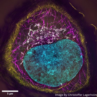
The purpose of this course is to introduce course participants to above techniques, first in theoretical lecture form, and second in practical form by a combination of instructor demonstrations and subsequent supervised practical work by course participants on provided samples including as shown above of fixed U2OS cells stained for DNA (DAPI; cyan), F-actin (Phalloidin-Alexa 488; yellow), microtubules (anti-α-tyrosinated tubulin/anti-IgG-Alexa 555; magenta), and mitochondria (anti-TOM20/anti-IgG-Alexa 647 ; grey).
Light Microscopy Techniques to be covered in the microscopy course:
- Zeiss Airyscan and Leica Lightning enhanced confocal microscopy (Zeiss 880/Leica Stellaris 8)
- Structural Illumination Microscopy (OMX SIM v4)
- Stimulated Emission Depletion Microscopy (Abberior)
- Single Molecule Localization Microscopy (Oxford Nanoimager)
The course will furthermore include lectures and practical demonstrations of image analysis tools that are applicable for analyzing images acquired within the course, and with image data archiving options in OMERO.
At the end of the course, the attendees will have gained a theoretical background of super-resolution techniques and will have gained invaluable practical experience with optimization of super-resolution microscopy techniques. Additionally, the attendees will have gained practical experience in the processing and analysis of images produced by these super-resolution microscopy techniques. This course will also provide a unique opportunity for networking and engaging in the Turku imaging community by making it easy for researchers to locate the experts and technologies and increase the efficiency and quality of the research.
Speakers and trainers
- Microscopy: Christoffer Lagerholm, Jouko Sandholm, Kaisa Rautaniemi, Elena Tcarenkova
- Image Analysis: Pasi Kankaanpää, Junel Solis
Application Information
Total course enrollment is 12 researchers. This course is suitable for researchers from Master Student level and up where researchers preferentially have prior experience with confocal microscopy and better yet a need/interest in exploring the benefits of super-resolution microscopy in their on-going research.
- Apply by: August 31st, 2024. Results will be published by September 3rd.
- Application Form: Click here to apply
Suggested Reading
- Wegel, E., Göhler, A., Lagerholm, B. et al. Imaging cellular structures in super-resolution with SIM, STED and Localisation Microscopy: A practical comparison. Sci Rep 6, 27290 (2016). https://doi.org/10.1038/srep27290
Course Organizers
- Cell Imaging and Cytometry Core, Turku Bioscience Centre, Åbo Akademi University & University of Turku, Turku, Finland.
- Turku BioImaging, Åbo Akademi University & University of Turku, Turku, Finland
For further questions, please contact:
- Christoffer Lagerholm, christoffer.lagerholm@abo.fi
- Irina Belaia, irina.belaia@abo.fi
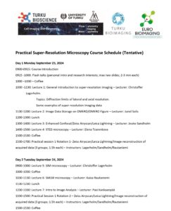
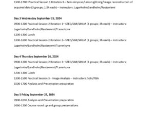
Tentative Schedule