The programme has been assembled on the true imaging strongholds of Turku and comprises a truly interdisciplinary array of prominent research groups and departments to share advanced imaging techniques in
- basic cell and molecular biology
- in disease characterization at the molecular level
- in patient diagnostics
The two-year programme is jointly administered and run by the two universities in Turku, the University of Turku and the Åbo Akademi University. The programme is administered by the Faculty of Science and Engineering at Åbo Akademi University and the Faculty of Medicine at the University of Turku. Successful completion of this two-year full-time programme results in the award of a Master of Science (M.Sc.) degree (Swe: Filosofie magisterexamen) 120 ECTS.
Both Finnish and foreign students who have completed a lower university degree equivalent to a Finnish B.Sc. degree are welcome to apply for the programme. The education is given in English.
The tuition fee is 12 000€/year for students applying outside the EU/EEA -area. Scholarships are available. No tuition fee for European students.

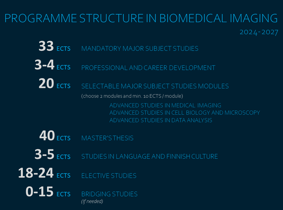
The studies consist of mandatory courses in biology, physics, engineering, microscopic applications, image processing and ethics. They aim to give the student the required basic knowledge in the field of biomedical imaging.
Students can select elective courses from a variety of applicable topics according to their interests.
The programme curriculum covers the following topics:
- Light microscopy, basic and advanced techniques
- Electron microscopy
- Tissue imaging and histopathology
- Nuclear medicine and magnetic resonance in imaging
- Tomographic imaging techniques
- In vivo non-invasive imaging
- Nanotechnology
- Imaging probes for light microscopic and nuclear imaging modalities
- Bioimage informatics and use of various image analysis tools
- Data analysis methods, such as machine learning and artificial intelligence
See Biomedical Imaging course structure for detailed information about the mandatory and selectable courses.
Application for the next academic year will be open 8.1. – 22.1.2025.
The MSc Programme in Biomedical Imaging is jointly administered by Department of Biosciences at Åbo Akademi University and the Faculty of Medicine at the University of Turku. In order to increase the chances of being admitted, applicants are advised to apply to both Universities.
The eligible applicants should have sufficient background knowledge in medical, biological and/or natural sciences, and they should have completed a lower university degree equivalent to a Finnish B.Sc. degree in the Life Sciences or in a relevant field (biomedical sciences, physics, engineering or chemistry).
Applications for the programme are evaluated by the admission committee. The admission committee scores the applicants according to their background and selects maximum of 50 applicants for the interview round.
No tuition fee for European students. The tuition fee is 12 000€/year for students applying outside the EU/EEA -area. Scholarships are available.
APPLYING STEP-BY-STEP:
1. Please read the admission instructions of the universities carefully:
Åbo Akademi, University of Turku.
2. Book and take the language test in advance if needed.
All applicants to the International Master’s Programmes taught in English must always prove their level of proficiency in the English language. Please find more information about language requirements in Åbo Akademi and the University of Turku.
3. Application through Studyinfo portal to both Åbo Akademi University and University of Turku.
Fill in the required information with care. Please note that to apply to both universities you must complete an online application to both universities and send in two sets of enclosures.
In the online application form, it is enough that you attach your enclosures electronically. In case one of the universities need the hard copies, they will contact you personally.
4. Submit the application and send all the required enclosures.
The application has to be admitted before January 22nd 2025 3.00pm.
Possibilities after graduation
After the studies students can work in several fields in Finland or abroad. The students can select courses to support their own career goals.
Some career paths of our graduates:
- continue as postgraduate students to pursue career as scientist
- work in imaging core facility
- work in science administration nationally or internationally
- work in hospital research laboratory
- industry and industrial research
- popular science projects
Why to Choose Turku as your next city
Education from the top of the world
Finland is a Nordic welfare state where equality is the fundamental ideology behind education. International rankings agree with us, Finland is a great place to study. Finnish higher education consists of two complementary sectors: universities promoting research and academic education, and universities of applied sciences offering professional higher education with close ties to the working life. In Turku there is six higher education institutes.
Accommodation
Turku offers a wide range of housing options for students near the campuses and the centre. The price of housing is more student-friendly than in many other cities in Finland.
The Student Village Foundation of Turku (TYS) was founded by the Student Union of Turku University and it specially builds, renovates, maintains and rents apartments for students in the Turku area. TYS has around 7 300 apartment places and 19 housing locations. As these apartments are specially directed to students, the rent prices are affordable. All TYS apartment rents include water, electricity, Internet connection and sauna turns.
Getting around is easy and cheap
In Turku everything is close. Turku is a fantastic city for cycling. Distances from place to place are short and traffic design favours bikes. The vast majority of people who live in less than ten kilometers from the city center cycle on a daily basis to work and school. In addition, the movement is easy thanks to a comprehensive local transport.
With the valid student card you will automatically get a discount of bus and train tickets in Finland. One month bus card for the whole Turku area costs 33€ (for student) and it can be used unlimitedly.
Student lunches
As the university students you will have an opportunity to offered government-subsidised student lunches for €2.60, which usually includes the main dish, side salad, bread, water and a non-alcoholic drink (e.g. milk or juice). Students are expected to show a valid student card each time to get the student-priced lunch. You can find dozens of student cafeterias around the campus area.
Freely available courses
As a student of the University of Turku or Åbo Akademi you have a right to take any courses provided by the universities freely. Both universities provide high quality education in English language in many fields of study. There is also a great possibility to study various languages in the university’s language centre. Some limitations may occur with some of the practical courses where the amount of participants is more limited.
Healthcare
With a valid Finnish university student card you have the right to free Finnish Student Health Service (FSHS). The health services provided are general practitioners and specialists, nursing care and physiotherapy as well as x-ray and laboratory work. FSHS also provides assistance in managing yourself during your studies. FSHS mental health services include mental health counselling, psychotherapy and psychiatric care. Dental care is also included to FSHS and fees are from 16€ to max. 30€.
For the special care such as hospital care and some specialist services, students who have Turku as their place of residence have the right to municipal health care.
Outdoor wonders
Turku is city by the sea. The Turku Archipelago has more islands than any other archipelago in the world. It has also been described as the world’s most beautiful archipelago. In addition there is three other national parks in Southwest Finland. Nature in near, it’s clean and magnificent – and it can be reached easily.
City of students got it all
Turku offers plenty of historical attractions, contemporary art and incredible nature. Explore the city’s many restaurants, shops, bars and other free time activities. As a popular student city you get to meet people from different cultures all over the world. Student culture is active: 20 percent of Turku residents are higher education students. People of Turku, “turkulaiset”, are warm hearted, open minded and kind people.
Student experiences
Toiba, India
(started 2018)"Now when studying in the Biomedical Imaging program, I am even more convinced that this is the right path for me."
Imran, Pakistan
(started 2015)"BIMA challenged me to step out of my comfort zone by and helped me to think outside the box. Without this program, I wouldn’t be living my dream of doing a doctorate in Neurology."
Leon, Estonia
(started 2016)"BIMA is an exciting opportunity to go beyond the familiar limits of your field and be at the forefront of the modern scientific research."
Hanna, Finland
(started 2017)
"Earlier I have studied Biotechnology. From the BIMA Program, I get the crucial expertise to my career and in addition, I am able to see the invisible."
Dado,Bosnia and Herzegovina
(started 2018)"It's been a wonderful experience and I love being in Turku. Just go for it and don’t be afraid of the unknown."
Sadaf, Iran
(started 2019)"Even though we have different academic backgrounds, I have decided to follow my sister, who also graduated from Biomedical Imaging few years ago. This proved to be a great decision, and I have no regrets making this life choice”
Programme Graduates
Joanna Pylvänäinen
“Lymphatic and vascular endothelium as barriers for transendothelial migration of breast cancer cells”
Petra Miikkulainen
“The effect of HIF prolyl hydroxylase PHD3 on activating migration and growth of carcinoma cells”
Mueez U Din
“Differentiation of Metabolically Distinct Areas within Head and Neck Region using Dynamic 18F-FDG Positron Emission Tomography Imaging”
Yaqi Qui
“Dietary factors affecting human aromatase gene reporter expression in normal tissues and cancer; focus on mammary gland and breast cancer”
Daniel Ebot
“The role of keratin intermediate filaments for the function of pancreatic islets and type 1 diabetes”
Yonas Mekuria
“Clinical Application of of Electronic Portal Imaging Device (EPID) and its quality control”
Mohamed El Missiry
“Combination of leukocyte phenotype with phosphoflow and cell size in the study of hematological conditions.”
Mehrbod Mohammadian
“Anatomical references and image analysis software in small animal brain imaging with PET tracers”
Muhammad Arsalan Khan
“Rotating Frame Relaxation Measurements in Experimental Acute Phase Cardiac Infarct in vivo”
Mojtaba Jafari Tadi
“An Accelerometer-Based Method for Simultaneous Extraction of Respiratory and Cardiac Gating Signals for Nuclear Medicine Imaging.”
Bettina Hutz
“Protein interactions in mitochondrial nucleoids before and after the knock-down of the mitochondrial inner membrane protein ATAD3”
Sanaz Nazari Farsani
“Precision and Accuracy of Marker-Based and Model-Based Radiostereometric Analyses in Determination of Three Dimensional Micromotion of a Hip Stem”
Basit Iqbal Butt
“Stromal interaction molecule 1 and its role in the regulation, proliferation and protein expression in follicular ML-1 thyroid cancer cells”
Behnoush Foroutan
“Myocardial free fatty acid uptake before and after bariatric surgery induced weight loss”
Dareen Mustafa Fteita
“Ultrastructural changes of Fusobacterium nucleatum as a defense mechanism against human neutrophil peptide-1 – A Transmission Electron Microscopy (TEM) study”
Prince Yaw Dadson
“Effects of bariatric Surgery on Abdominal Adipose Tissue Distribution in Severely Obese Patients”
Shereif Haykal
“Evaluation of Transcatheter Aortic Implantation candidates using Computed Tomography”
Elena Tcarenkova
“Developing Correlative Optical-Photoacoustic Microscopy for high-resolution in vivo imaging”
Natalia Gurvits
“Optimization of immunofluorescence protocols for detection of biomarkers in colorectal and breast cancer tissues”
Guru Prasad Padmasola
“Exploring the feasibility of Al18F-nota folate tracer for assessment of atherosclerotic plaques in mice”
Ezgi Özliseli
“Development of novel imaging probes and related methods for cellular labeling and tracking”
Kai-Lan Lin
“A Study on the Recycling Mechanism of Dap160, a Scaffolding Protein in the Neuromuscular Junction of/Drosophila melanogaster/ Larvae”
Sina Tadayon
"In Vivo Imaging of Experimental Animal Model of MS Using Novel VAP-1 Targeting Radiotracer”
Marjukka Elmeranta
“Stimulated emission depletion microscopy of sub-diffraction polymerized structures fabricated with two-beam direct laser writing lithography”
Kaveh Nik Jamal
“Three-dimentional fourier analysis of electron microscopy to characterize diffusion tensor imaging data”
Prakirth Govardhanam Narayana
“Development of nanoantibiotics and evaluation via in vitro and in vivo imaging”
Tatsiana Auchynnikava
“68Ga-NOTA-Siglec-9 – a new Vascular Adhesion Protein-1 targeted agent for inflammation imaging”
Jaakko Suominen
“3D printed UV-curable polydimethylsiloxane scaffolds loaded with bovine serum albumin”
Kumail Motiani
"Two weeks of training reduces liver fat content and improves liver substrate uptake and lipoprotein subclasses"
Anastasiia Driuchina
"Association between intrathoracic adipose tissue mass and metabolic parameters"
Maxwell Miner
"Semi-automated Synthesis of High Purity (2S, 4R)-4-[18F]fluoroglutamine and its Use for PET Imaging Experimental Gliomas"
Kalpana Parajuli
"Creating a CT Template-based Attenuation Correction Method for Pelvic PET/MRI Imaging"
Svetlana Bezukladova
"Multimodal approach to the evaluation of diffuse neuroinflammation in multiple sclerosis using positron emission tomography and diffusion tensor imaging"
Ciaran Butler-Hallissey
"Uncovering the LINC between cytoplasmic keratin 8 and nuclear lamins using 2D and 3D culture systems"
Md Rashedur Rahman
"Identification of glycosylations affected by androgen-regulated process on the surface of extracellular vesicles derived from PCa cell lines"
Aruna Ghimire
"Colonocyte keratins are up-regulated in hypoxia and affects the nuclear translocation of HIF-1a"
Sami Şanlıdağ
"3D Bioprinting of Nanoparticle Incorporated Cell-Laden Bioink for Muscle Tissue Engineering"
Allison McGuire
"Adapting a Fluorescence-Based Superoxide Detection Method in order to Visualize ROS in the Foliar Mitochondria of Arabidopsis thaliana"
Angeli Kumari
"Keratins in β-cells of the pancreas and their role for mitochondria and insulin vesicles"
Nuria Barber-Perez
"Polyacrylamide Stiffness-gradient Hydrogels: A 2D Culture System to Study Cell Mechanoresponsiveness to Substrate Stiffness"
Erika Atencio Herre
"Radiosynthesis and biological evaluation of [68Ga]Ga-NOTA-Folate in experimental atherosclerosis and myocardial infarction models"
Sabrina Benoit
"Fluorescence imaging of Drosophila melanogaster tracheal system: investigating the morphology of LUBEL mutant flies"
Defne Dinç
"Quantitative Analysis of Force Transduction in Human Mammary Epithelial Cell Populations"
Tzu-Chen Rautio
"Effects of Nanomaterial-Delivered Protein Kinase A on Human Breast Cancer Stem Cells"
Vincensa Nicko Widjaja
"Developing an in vitro model and quantitation method for bone marrow adiposity research"
Sujan Ghimire
Characterisation of cancer cell adhesion on endothelial cells by super-resolution microscopy
Brajesh Shrestha
Synthesis of CuS@MSN@SpAcDex for Tacrolimus delivery to treat End Stage Renal Disease
Thalla Divyendu Goud
Development of a rotatory device for development of 3D super-resolution tomographic STED microscopy
Doroteja Bednjanec
Targeted Sequence Delivery Platform with ROS-Response for the Treatment of Osteoarthritis
Anna Jalo
Hyperinsulinemic euglycemic clamp in anesthetized rat: A protocol for rat in vivo FDG PET studies
Rasha Elmansuri
Visualization of cellular surface and intercellular structures of human pluripotent stem cells using electron microscopy techniques
Seyed Hosseini
Applying Transfer Learning in Classification of Ischemia from Myocardial Polar Maps in PET Cardiac Perfusion Imaging
Shaoxia Wang
The functional role of NOTCH and potential NOTCH associated genes in head and neck cancer
Yasaman Anbari Yazbi
"A quantitative and qualitative analysis of Positron Emission Tomography (PET) in Yttrium-90 radioembolization; investigating the utility of PET dosimetry in identifying sites of necrosis and viable tumor"
Fariha Annesha
"A Framework for the Systematic Comparison of Classical Non-Learning, Machine Learning, and Artificial Intelligence Approaches to Cell Segmentation & Nuclei Detection in Histopathological Images"
Rafael Diaz
"Review of the CH-STED performance and application range over conventional STED and z-STED"
Heini Hautanen
"Quantification of hepatic steatosis and fibrosis in pre-clinical mouse models by applying digital image analysis"
Petar Kosijer
"The role of b-cell keratins in islet histology, glucose uptake, and diabetes susceptibility"
Shiva Latifi
"Effect of breast cancer, doxorubicin chemotherapy and exercise on heart vascularization and cardiomyofibers"
Jessica Salo
"Intracellular Delivery OF CRISPR/Cas9 Plasmid With Zif-8 Nanocarrier to Liver Cancer Cells"
Maria Tarczewska
"Visualizing intra- and extracellular vesicles of B cells using expansion microscopy"
